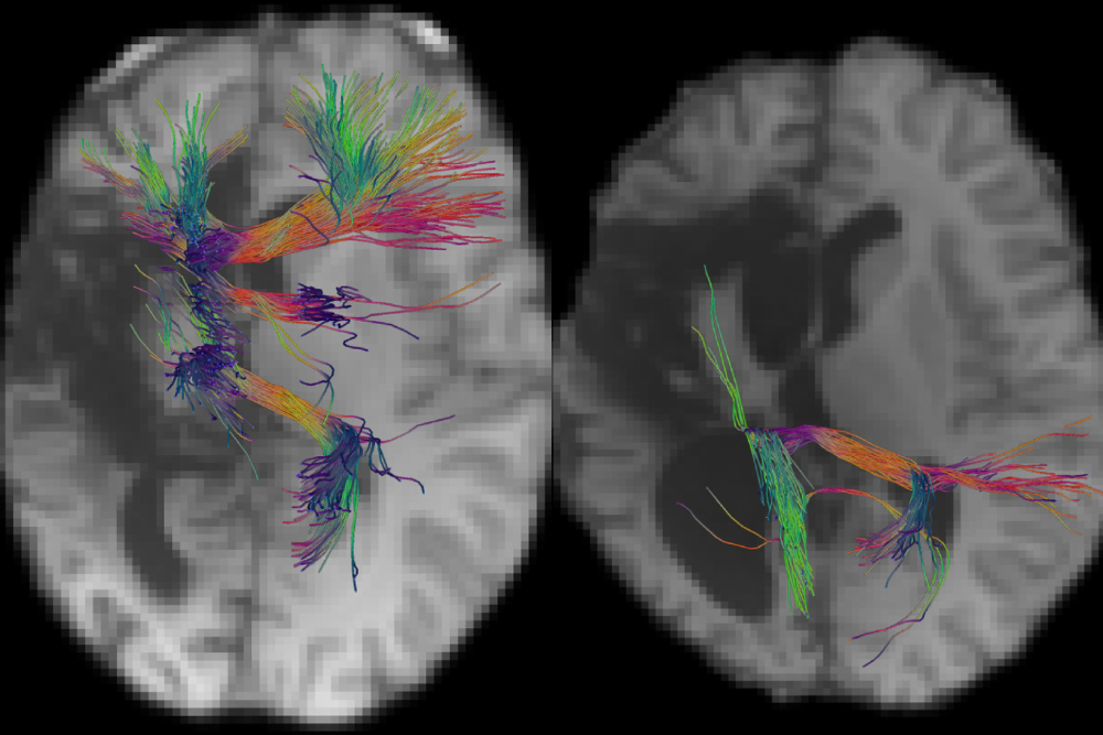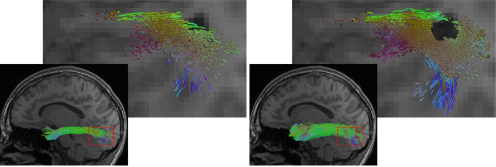
How can nerve pathways in the brain be visualized to improve the planning of complex surgeries? A research team from the Lamarr Institute and the University of Bonn, in collaboration with the Translational Neuroimaging Group at the Departments of Neuroradiology and Epileptology at the University Hospital Bonn (UKB), has investigated an AI-powered method that makes these reconstructions more precise. The study, recently published in NeuroImage: Clinical, could ultimately help make neurosurgical procedures safer.
What Is Tractography – and Why Is It Important?
The brain is a highly complex network of nerve cells interconnected by delicate pathways – known as nerve fibers or tracts. These connections are essential for movement, speech, thought, and many other functions. To visualize these structures, researchers use tractography, an imaging technique that calculates the course of nerve pathways based on specialized MRI scans. This information is particularly crucial for planning brain surgeries, such as those performed on epilepsy patients undergoing surgical intervention.
Current tractography methods rely on mathematical models that infer the location of nerve pathways from MRI data. However, these methods often involve uncertainties, especially when the brain has been altered due to disease or surgery. This is where modern AI methods come into play: By leveraging machine learning, these systems can recognize patterns and generate more accurate reconstructions.

AI-Powered Tractography Shows Potential – But Also Challenges
In the study, the researchers tested a widely used AI method called TractSeg, originally trained on healthy brains. The team investigated whether it could also work for epilepsy patients who had undergone a hemispherotomy – a surgical procedure that disconnects the two hemispheres of the brain.
The results showed that TractSeg performed well in many cases but also produced unexpected errors: It reconstructed nerve pathways that should no longer exist due to the surgery – a phenomenon known as „hallucination.“ At the same time, some remaining pathways were either incompletely captured or entirely missing from the reconstruction.
A New Hybrid Approach for More Accurate Reconstructions
To address these issues, the team developed a new hybrid method that combines the advantages of AI with the data fidelity of traditional techniques. This approach ensures that only existing nerve connections are reconstructed. The result: No more hallucinations, better detection of preserved pathways, and overall more accurate reconstructions – even in healthy brains.
Prof. Dr. Thomas Schultz, Principal Investigator in the Life Sciences at the Lamarr Institute and a professor at the Institute for Computer Science at the University of Bonn, highlights the significance of this work:
„Our study demonstrates both the potential and the limitations of AI-powered tractography in clinical applications. Combining AI with traditional methods offers a promising solution for more precise reconstructions, especially when dealing with patient data affected by pathological changes. Our goal is to further refine these approaches and make them applicable for neurosurgery in the long run.“

Funded by a TRA Research Award from the University of Bonn
This publication is the result of a collaboration funded by the Modeling for Life and Health research award from the University of Bonn’s Transdisciplinary Research Areas (TRA) Modeling and Life & Health. The project was conducted in partnership with PD Dr. Theodor Rüber, Principal Investigator of the Translational Neuroimaging Group and a neurologist at the UKB’s Department of Neuroradiology.
The University of Bonn’s Transdisciplinary Research Areas (TRA) serve as innovation and exploration hubs in research and education, enabling scholars to collaborate across disciplines and faculties, as well as with external partners, to tackle key scientific, technological, and societal challenges. This first joint publication illustrates how the exchange between AI research and neuroscience can help advance medical procedures – offering direct benefits for patients.
Additionally, the project was supported by the BNTrAinee program at the University of Bonn and the Neuro-aCSis Bonn Neuroscience Clinician Scientist Program.
Publication
Grün, Bauer, Rüber, Schultz: Deep Learning Based Tractography With TractSeg in Patients With Hemispherotomy: Evaluation and Refinement, NeuroImage: Clinical. https://doi.org/10.1016/j.nicl.2025.103738
